znajdziesz nas na:


znajdziesz nas na:


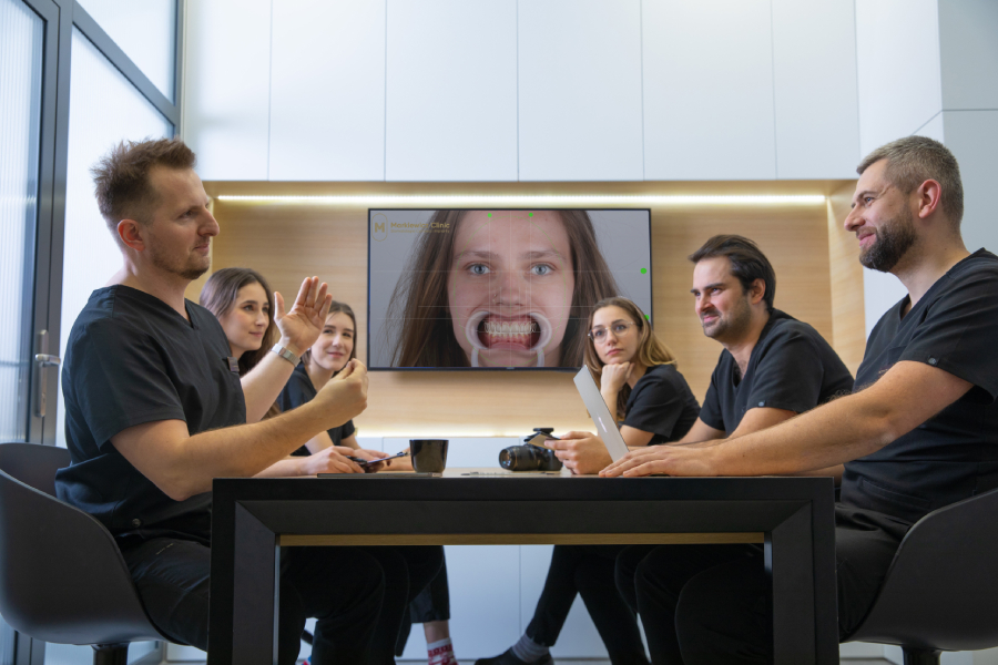
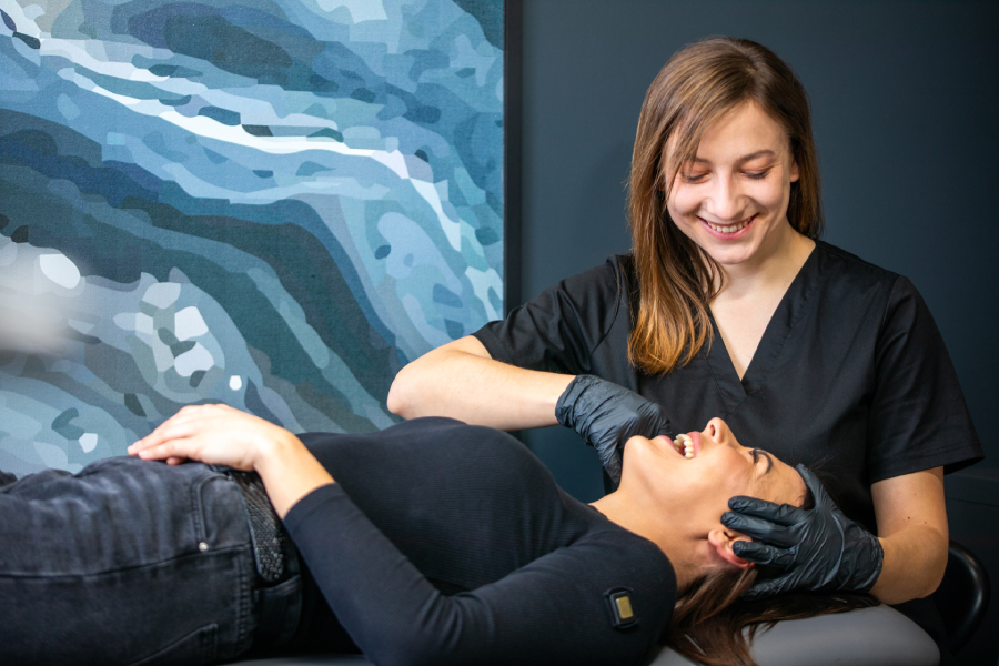
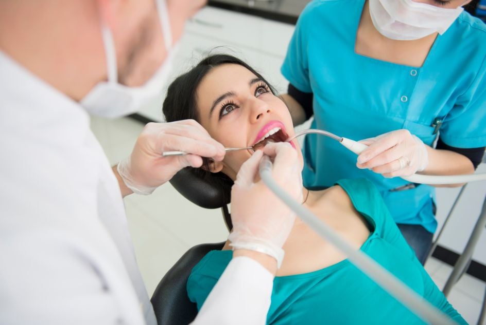
Modern dentistry offers many options for managing partial edentulism. One of the most commonly used solutions is a dental implant. However, in the region of the maxillary premolars and molars, a key challenge often arises—the close proximity of the maxillary sinus floor. When resorption of the maxillary alveolar process occurs, a maxillary sinus floor elevation may be required. This implantologic procedure enables the placement of dental implants even in compromised bone conditions.
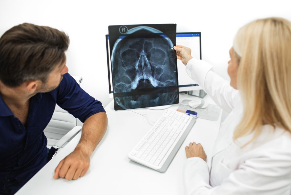
The maxillary sinus is an air-filled cavity within the maxilla, closely connected to the nasal cavity. Its inferior boundary—the sinus floor—often lies very near the roots of the maxillary premolars and molars. After tooth loss in this area, progressive alveolar bone resorption reduces the height of the alveolar ridge. Insufficient vertical bone height prevents stable placement of endosseous dental implants without encroaching on the sinus.
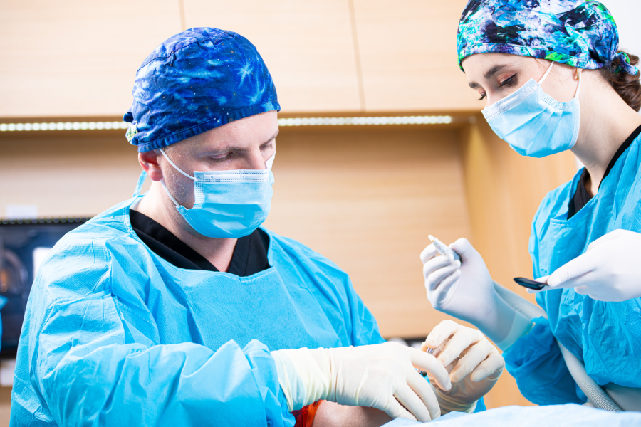
A maxillary sinus floor elevation—also known as a sinus lift—is a surgical procedure in which the Schneiderian membrane (sinus mucosa) is carefully separated and a bone graft (bone substitute material) is placed. This allows reconstruction of the alveolar bone and creates the conditions necessary for subsequent implant placement.
Two main techniques are distinguished:
The procedure proceeds in several steps:
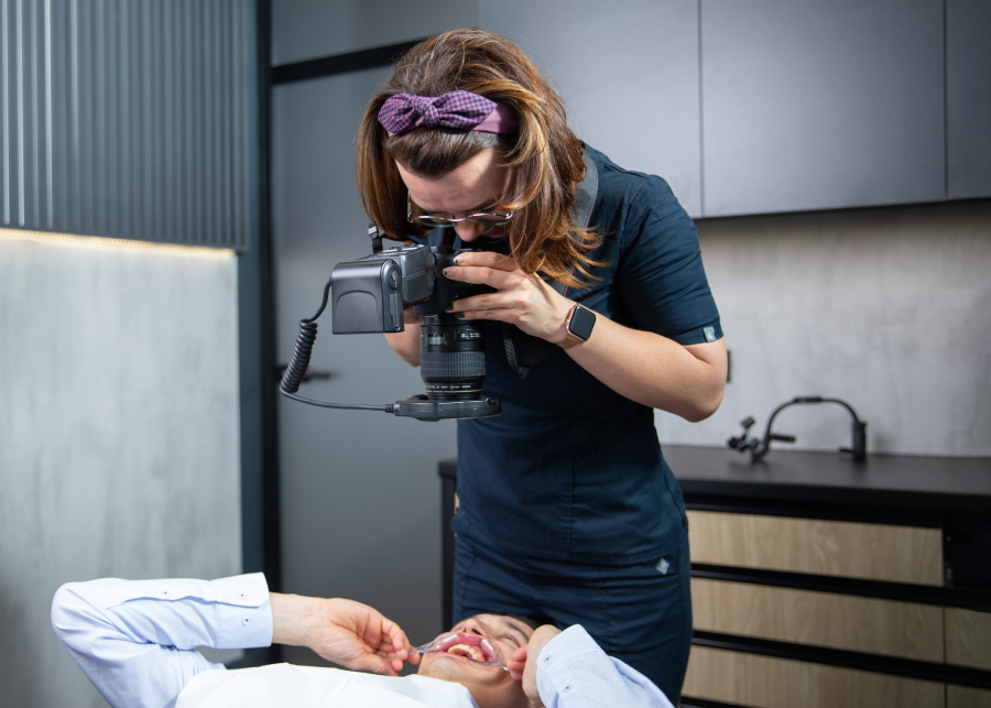
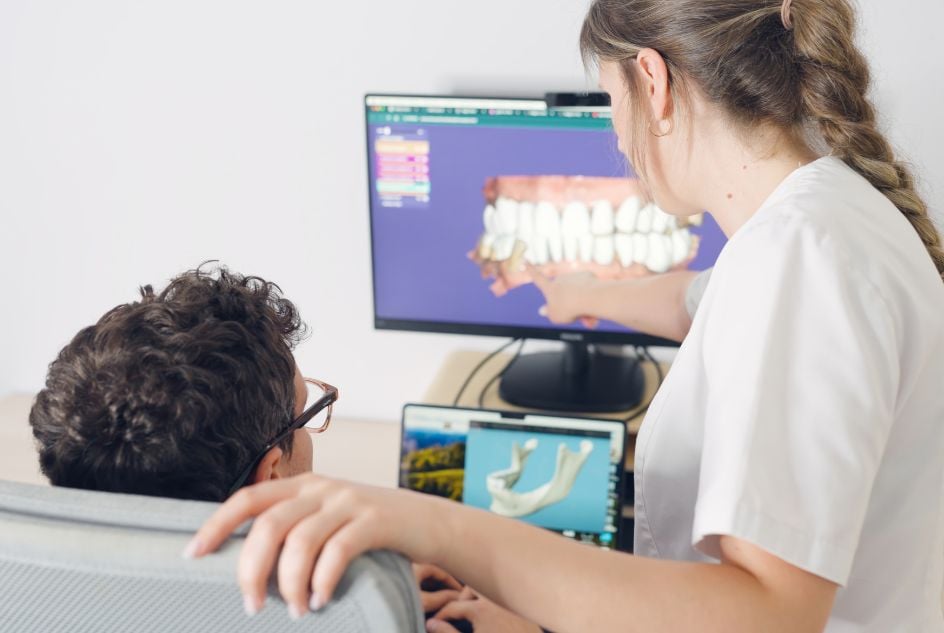
Implant placement and sinus floor elevation are often closely related. Under favourable bone conditions, both procedures can be performed during a single surgical session. This approach shortens the overall implant treatment time, allowing patients to restore missing teeth sooner. However, this is not always feasible—the final decision rests with the dentist, who evaluates the patient’s oral status and alveolar bone volume based on CT imaging.
If the vertical bone height in the maxillary alveolar ridge is insufficient for primary implant stability, implant placement must be deferred. In such cases, the surgeon first performs a sinus floor elevation and inserts a bone graft material that gradually integrates with the patient’s bone. The reconstruction process requires time—typically 6 to 9 months—for the augmented bone to mature fully and provide a stable foundation for future implants.
This protocol reduces the risk of complications, including migration of an implant into the sinus cavity, and significantly improves the overall success of implant therapy. Although it extends the time to final prosthetic restoration, it ensures safe implant placement and a durable aesthetic and functional outcome.
As with any surgical procedure, sinus floor elevation carries certain risks. The most common complications include:
Contraindications include, among others, chronic rhinosinusitis, uncontrolled systemic diseases, inability to maintain adequate oral hygiene, or persistently unfavourable bone conditions despite planned augmentation.
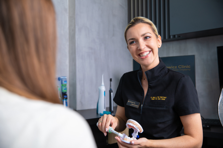
After surgery, patients should follow their dentist’s instructions:
Healing typically takes several months and is closely monitored by the dentist.
Sinus floor elevation is a key procedure in contemporary implantology, enabling treatment even in cases of advanced bone resorption. It restores adequate bone volume for dental implant placement in patients who have long struggled with missing teeth. Reconstruction of the maxillary alveolar ridge and increased bone height beneath the sinus allow the dentist to perform implant placement safely, minimizing complications and improving long-term prognosis.
Modern dentistry offers both the open (lateral window) and the less invasive closed (crestal/transalveolar) sinus lift techniques. The choice depends on the degree of bone loss, the amount of augmentation needed for implant stability, and the patient’s individual anatomy. In both approaches, the primary goal is to prepare the site for future implants that not only replace missing teeth but also restore proper masticatory function.
When performed by an experienced oral and maxillofacial surgeon or implantologist, a sinus lift is a safe and predictable procedure. Although it demands precision and care, implant outcomes after such preparation are durable, and dental implants can serve the patient for many years, ensuring both an aesthetic smile and everyday comfort. Importantly, appropriate diagnostics (CT imaging) and strict adherence to postoperative instructions maximise treatment efficacy and reduce the risk of complications such as rhinosinusitis or perforation of the Schneiderian membrane.
Read also: Bonding or Veneers


Uśmiechaj się pewniej każdego dnia
Odkryj, jak nowoczesne technologie i innowacyjne metody leczenia mogą przynieść rewolucję w dbaniu o Twoje zęby. Nasz zespół specjalistów jest gotowy wprowadzić Cię w przyszłość stomatologii
Markiewicz Clinic
ul. Karola Szymanowskiego 2 lok. 6
80-280 Gdańsk
+48 58 558 80 57
rejestracja@markiewiczclinic.com
znajdziesz nas na:

 Unia Europejska


 osobiście w rejestracji Kliniki,